Tissue characterization
Click on the toggle bars to read more:
Synchrotron X-ray tomography is a non-destructive method for 3D visualization and quantitative analysis of materials. In conventional X-ray tomography the contrast is given by the materials X-ray absorbance, consequently imaging soft materials can be extremely difficult. Phase-contrast Synchrotron X-ray tomography (SR-PhC-μCT) emerged more recently to image materials that weakly absorb X-rays and/or have similar densities. In SR-PhC-μCT the contrast is enhanced by the phase shifts that occur at the boundaries between materials with different refractive indexes, allowing the study of soft tissue structures at high resolution.
Our aim is to achieve a complete structural characterization of musculoskeletal tissues at the meso/microscale. 3D high resolution structural studies are fundamental to understand the complex function-structure relation. However, 3D imaging techniques suitable to unveil the microstructural organization of weakly mineralized (e.g. forming bones at early embryonal stages) and soft tissues (e.g. tendons and ligaments) have only recently emerged, and many studies were, until now, based on 2D histology.
Our research mostly focuses on the study of Achilles tendons, embryonic bone mineralization and how muscular contractions/loading affect them (see “bone mechanics”, “tendon mechanobiology”, or Connective knee joint tissue mechanics" for details). SR-PhC-μCT produces high-quality images that capture the complex 3D organization of soft and weakly mineralized tissues in conditions as close as possible to natural. Furthermore, SR-PhC-μCT can be used for in situ characterization of how soft tissue responds to mechanical stimuli on a microscopic level.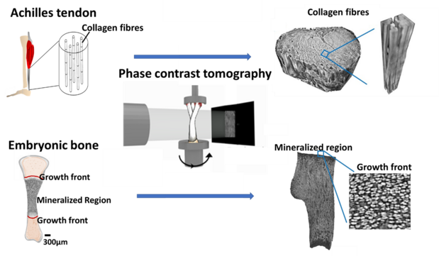
Figure: Phase-contrast Synchrotron X-ray tomography imaging is used to study the 3D microstructure of soft tissue such as in Achilles tendons and in still weakly mineralized embryonic bones.
Researchers at LU: Researcher: Maria Pierantoni; PI: Hanna Isaksson
Funding: Knut and Alice Wallenberg KAW Foundation, Carl Trygger Foundation, Kungliga Fysiografiska Sällskapet i Lund
Recent publications (external links):
- Pierantoni, Le Cann, Sotiriou, Ahmed, Bodey, Jerjen, Nowlan, Isaksson: Muscular loading affects the 3D structure of both the mineralized rudiment and growth plate at early stages of bone formation, Bone, 2021 doi.org/10.1016/j.bone.2021.115849
Pierantoni, Hammerman, Silva Barreto, Larsson, Notermans, Bodey, Eliasson, Isaksson, Spatiotemporal and microstructural characterization of heterotopic ossification in healing rat Achilles tendons. The FASEB Journal 37.6, 2023 https://doi.org/10.1096/fj.202201018RRR
Pierantoni, Hammerman, Silva Barreto, Andersson, Novak, Isaksson, Eliasson, Heterotopic mineral deposits in intact rat Achilles tendons are characterized by a unique fiber-like structure. Journal of Structural Biology: X 7: 100087, 2023 https://doi.org/10.1016/j.yjsbx.2023.100087
Musculoskeletal tissues are collagen-based materials and have a well-defined, intricate structure from the molecular building blocks up to the organ scale. The mechanical properties of these tissues depend on all length scales and the relation between them. The tissues are dynamic and adapts to their local loading environment. Thus, despite being very similar at the material scale, they are structurally different and exhibit different mechanical properties.
Our aim is to understand the relationship between mechanical stimuli and the basic building blocks of musculoskeletal tissues, mainly bone and tendon (see section on “tendon mechanobiology”). To do this, we need to understand how the structure and composition of these tissues are affected and responds to mechanical stimuli at the nanoscale, as well as how this relates to other length scales.
To study the structure of collagen and mineral, we are using high-resolution synchrotron techniques such as small- and wide-angle X-ray scattering as well as conventional techniques such as polarized light microscopy (PLM). To study the nanoscale composition, we are using high-resolution synchrotron techniques such as X-ray fluorescence, which probes the elemental content and conventional techniques such as Fourier-transform infrared spectroscopy, which probes the molecular content. In order to further unravel the relationship between mechanical stimuli, structure, composition and length scales, we combine all these methods with mechanical loading in vivo or in situ.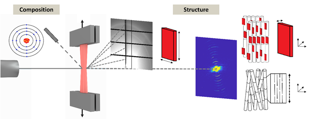
Figure: Schematic of synchrotron techniques we combine to study the structure and composition of musculoskeletal tissues. Information about the elemental composition is extracted from characteristic secondary X-rays released by excited atoms. Information of the nanoscale structure, arrangement and alignment are extracted from scattered X-rays at wide and small angles.
Researchers at LU: PhD student: Isabella Silva Barreto; PI: Hanna Isaksson
Funding: Knut and Alice Wallenberg KAW Foundation and Carl Trygger Foundation
Recent publications (external links):
- Silva Barreto, Le Cann, Ahmed, Sotiriou, Turunen, Johansson, Rodriguez‐Fernandez, Grünewald, Liebi, Nowlan, Isaksson: Multiscale Characterization of Embryonic Long Bone Mineralization in Mice, Advanced Science, 2020 doi.org/10.1002/advs.202002524
Pierantoni, Hammerman, Silva Barreto, Andersson, Novak, Isaksson, Eliasson, Heterotopic mineral deposits in intact rat Achilles tendons are characterized by a unique fiber-like structure. Journal of Structural Biology: X 7: 100087, 2023 https://doi.org/10.1016/j.yjsbx.2023.100087
Dejea, Raina, Silva Barreto, Sharma, Liu, Ferreira Sanchez, Johansson, Isaksson, Multi-scale characterization of the spatio-temporal interplay between elemental composition, mineral deposition and remodelling in bone fracture healing. Acta Biomaterialia, 2023. https://doi.org/10.1016/j.actbio.2023.06.031
Skeletal tissues, such as bone, tendon, and cartilage, are hierarchically structured composite materials. The arrangement of the building blocks at the nano scale, namely collagen type-I and hydroxyapatite mineral (for bone), and the structures they form on longer length scales, contribute to making the tissues optimized to withstand mechanical loading. X-ray techniques are state-of-the-art in skeletal research but has limitations in terms of contrast when applied to soft tissues, and when metallic implants are present. Neutrons interact differently with materials as compared to X-rays and are frequently used as a complementary probe in other fields of research.
Our aim is to investigate if neutron techniques can help us improve the understanding of how degenerative disorders such as osteoarthritis (OA) and trauma treatments such as medical interventions involving metallic implants affect the composition and structure of skeletal tissues.
The project investigates possible applications for neutron techniques such as small-angle neutron scattering (SANS) and neutron tomography (NT), and compare the results to respective X-ray equivalent, i.e., small-angle X-ray scattering (SAXS) and X-ray tomography (XRT).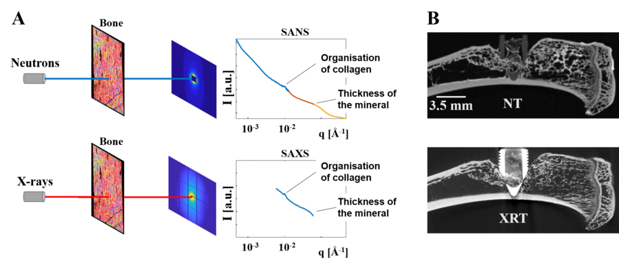
Figure (A) Small-angle neutron and X-ray scattering (SANS and SAXS) from bone, characterizing the nano structural building blocks. (B) Neutron and X-rat tomography (NT and XRT) of skeletal tissues and a metallic implant.
Researchers at BME: PhD student: Edvin Tobias Bokvist Wrammerfors; PI: Hanna Isaksson
Funding: Swedish Foundation for Strategic Research through the SwedNess graduate school.
Recent publications (external links):
- Törnquist, Le Cann, Tengattini, Helfen, Kok, Hall, Isaksson. The hydration state of bone tissue affects contrast in neutron tomographic images. Frontiers in Bioengineering and Biotechnology, section Biomaterials 10:911866, 2022 doi.org10.3389/fbioe.2022.911866
- Törnquist, Le Cann, Tudisco, Tengattini, Ando, Lenoir, Hektor, Raina, Tägil, Hall, Isaksson. Dual modality neutron and X-ray tomography for enhanced image analysis of the bone-metal interface. Physics in Medicine and Biology 66, 135016, 2021 doi.org10.1088/1361-6560/ac02d4
- Törnquist, Gentile, Prévost, Diaz, Olsson, Isaksson: Comparison of small-angle neutron and X-ray scattering for studying cortical bone nanostructure, Scientific Reports, 2020 doi.org/10.1038/s41598-020-71190-9