Connective knee joint tissue mechanics
Mechanobiological modelling of osteoarthritis progression
Articular cartilage is a soft tissue that covers the diarthrodial joints. Its major components are collagen type II, proteoglycans, chondrocytes, and water. Osteoarthritis (OA) is a common musculoskeletal disease that degenerates the articular cartilage affecting over 50 million Europeans. Since the progression of OA is highly patient-specific, prevention of the disease can only become possible when the progression can be estimated for an individual subject.
Our research also aims at characterizing how the mechanical properties of cartilage evolve as OA progresses, using Finite Element simulation and experimental data. This is combined with research about understanding the adaptive mechanobiological response of cartilage due to overloading caused by overweight or joint injury. Finite element modeling with patient-specific motion analysis and musculoskeletal modeling are combined to estimate the progression of knee OA and compare those predictions to clinical follow-up data.
Additionally, our research investigates novel techniques for characterizing ex vivo samples of cartilage and meniscus to investigate signs of OA development. We work with phase-contrast enhanced synchrotron tomography as well as neutron tomography to image cartilage and meniscus, allowing us to determine properties such as cell density, fiber orientation, or water diffusivity under different mechanical loads and/or different stages of OA development.
This project will help to comprehend how cartilage degeneration begins after trauma or higher tissue deformations due to overweight. The results will provide new information to validate our computational predictive models. These mechanobiological models will allow rapid and clinically feasible prediction of OA and simulation of the effect of interventions.
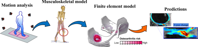
Figure: Workflow of the research project. Motion analysis of patients is conducted to determine the loads in the knee using musculoskeletal models. This data is implemented in FE models developed from MRI information. After the implementation of mechanobiological algorithms, numerical predictions of cartilage degeneration are compared to clinical follow-up data.
Researchers at LU: Postdoc: Anna Gustafsson; PhD students: Viktor Jönsson, Edvin Tobias Bokvist Wrammerfors; PI: Hanna Isaksson
Funding: Swedish Research Council (2019-00953 - under the frame of ERA PerMed), Novo Nordisk Foundation (grant no. NNF21OC0065373).
Recent publications (external links)
Orozco, Tanska, Gustafsson, Korhonen, Isaksson. Crack propagation in articular cartilage under cyclic loading using cohesive finite element modeling.” J Mech Behav Biomed Mater.(2022), 131, 105227 https://doi.org/10.1016/j.jmbbm.2022.105227
Orozco, Karjalainen, Moo, Stenroth, Tanska, Rios, Tuomainen, Nissi, Isaksson, Herzog, Korhonen, A musculoskeletal finite element model of rat knee joint for evaluating cartilage biomechanics during gait.” (2022)PLOS Comput Biol 18(6): e1009398. https://doi.org/10.1371/journal.pcbi.1009398
Orozco, Eskelinen, Kosonen, Tanaka, Yang, Link, Ma, Li, Grodzinsky, Korhonen, Tanska. “Shear strain and inflammation-induced fixed charge density loss in the knee joint following ACL injury and reconstruction: a computational study” Journal of Orthopaedic Research. (2021), https://doi.org/10.1002/jor.25177