Trapping
Cell/Particle trapping
Particles can be trapped and retained against flow by means of acoustic radiation forces. A common way of implementing an acoustic trap is to use a small piezoelectric transducer to generating a local acoustic standing wave field in a micro channel. Microparticles that enter the transducer region will be influenced by the primary l/2 standing wave field and focused in the pressure node plane (Fig 1a). At the same time the particles are also influenced by the lateral gradient field that force particles together in the pressure nodal plane (Fig 1 b-d). This mode of operation enables efficient enrichment of particles and cells and once trapped buffer conditions can be altered e.g. offering in situ labelling and washing.

Figure 1. Principle of acoustic cell/particle trapping in a l/2 standing wave.
Submicron particle trapping
Acoustic trapping works well under standard acoustophoresis conditions, i.e. when the radiation force on the particles is significantly larger than Stokes drag forces induced by acoustic streaming or perfusion flow. Consequently, acoustic trapping systems operated in the low MHz range display poor ability to trap submicron particles. However, if larger particles, so called seed-particles, 5-10 um, are pre-loaded in the trapping region, submicron particles will be attracted to the seed-particles by the inter particle forces that arise due to scattered sound interaction between the seed particles and the submicron particles, see figure below.
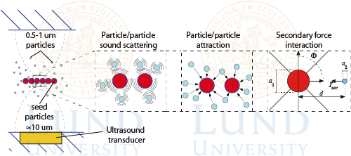
By preloading the trapping unit with seed-particles and aspirate e.g. a dilute bacteria suspension enrichment of pathogens can be accomplished in a short time period with minimal losses. More recently it has also been demonstrated that submicron bioparticles such as extracellular vesicles (EV) can be enriched and isolated and washed from its background matrix, enabling studies of biomarkers specifically linked to the EV population. The figure below shows a schematic of seed-particle mediated trapping of submicron particles. Most importantly, EV isolation can be accomplished from very small sample volumes (< 50 ul), enabling access to e.g. blood plasma or CSF biobanks.

References
Cell/Particle trapping
Lilliehorn T. et al, Dynamic arraying of microbeads for bioassays in microfluidic, Sensors and Actuators B: Chemical, 2005, 106, 2, 851-858
Evander M. et al., Non-invasive acoustic cell trapping in a microfluidic perfusion system for on-line bioassays, Analytical Chemistry, 2007, 79, 2984-2991
Norris JV et al., Acoustic Differential Extraction for Forensic Analysis of Sexual Assault Evidence, Analytical Chemistry, 2009, 81, 15, 6089-6095
Hammarström B. et al., Non-contact Acoustic Cell Trapping In Disposable Glass Capillaries, Lab Chip, 2010, 10, 17, 2251-2257
Tenje M. et al., Acoustic trapping as a generic non-contact incubation site for multiplex bead-based assays, Anal Chim Acta, 2015, 853, 682-688
Submicron Particle trapping
Hammarström B. et al., Seed particle enabled acoustic trapping of bacteria and submicron particles in continuous flow systems,Lab Chip, 2012, 12, 4296-4304
Evander M. et al., Non-contact acoustic capture of microparticles from small plasma volumes, Lab Chip, 2015, 15, 12, 2588-2596,
Ohlsson P. et al., Integrated acoustic separation, enrichment and microchip PCR detection of bacteria from blood for rapid sepsis diagnostics, Anal Chem, 2016, 88, 19, 9403-9411
Rezeli M. et al., Comparative proteomic analysis of extracellular vesicles isolated by acoustic trapping or differential centrifugation, Anal Chem, 2016, 88, 8577−8586
Ku A. et al.,Enrichment of Extracellular Vesicles from Biological Fluids, Anal Chem, 2018, 90, 8011-8019
Ku A. et al., A urinary extracellular vesicle microRNA biomarker discovery pipeline; from automated urinary extracellular vesicle enrichment by acoustic trapping to microRNA sequencing, 2019, PLOS ONE, 14(5), e0217507
Gidlöf O. et al., Proteomic profiling of extracellular vesicles reveals additional diagnostic biomarkers for myocardial infarction compared to plasma alone, Scientific Reports, 2019, 9, 8991