Our Research


Dolphins and other toothed whales use echolocation - a way of investigating their surroundings by emitting short ultrasound pulses termed “echolocation clicks”, which bounce back to the animals as reflections from objects in the water. Humans also use this pulse-echo principle in ultrasound machines and in other acoustic non-destructive testing devices. However, the animals’ ultrasound abilities far outscores the yet invented human ultrasound technology.
We study dolphin echolocation to better understand the pulse-echo based ultrasound strategies developed through millions of years of evolution, rendering them to be the most skillful echolocators on the planet. We also search for the mechanisms behind the production of the echolocation signals and investigate how we can implement this new knowledge in the pulse-echo based technology of tomorrow e. g. in future ultrasound machines for human diagnostics and other acoustic non-destructive material testing methods.
ELVIS - an EchoLocation and Visualization Interface System
The system can visualize the dolphin echolocation by interpreting the signals from an array of hydrophones and creates a video image representing the location and intensity of the dolphin sound. This video image can be viewed by the researcher. This makes it easier to understand how the dolphin uses its echolocation. Another very exciting feature is that the video image also can be shown to the dolphin in real-time, making it possible for the dolphins to draw computerized pictures for the first time in history!
By further developing this feature it is also possible to set up the system as an acoustically operated touch screen. Then the dolphins can use their quite narrow sonar beam to click on buttons on the screen in the same manner as we use the mouse cursor on a computer.
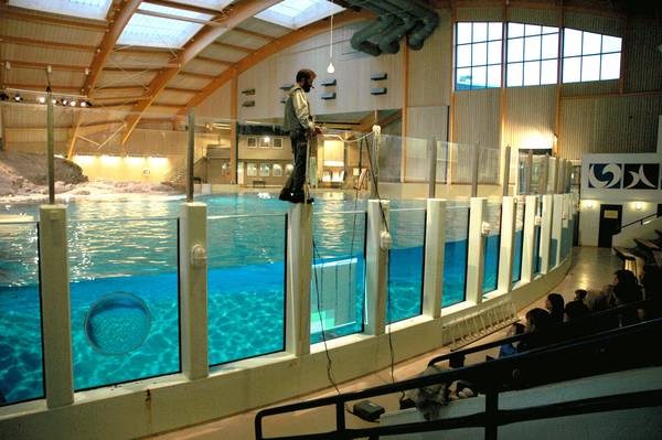
In the foto above from the Lagoon at Kolmården Wild Animal Park, the visual feedback of the dolphin's sonar beam is represented by a light intensity graph. You can see the dolphin echolocating at the lower corner of the screen.
Future medical imaging technology moves in the direction of molecular imaging where a contrast agent has been functionalized with tumor/tissue specific targeting agents (i.e. antibodies, affibodies, peptides, aptamers). Thus the contrast particle binds specifically to e.g. tumor tissue and can be enriched there. In other words, what is probed is the molecular abnormality that is the basis of the disease, rather than imaging the end effects or symptoms of these molecular alterations (such as anatomy or blood flow changes). One such promising contrast agent is nano-particles. However, nano-particles are too small to be visualized using conventional ultrasound techniques.
Inspired by a technique named magnetomotive optical coherence tomography it was suggested that a superparamagnetic iron oxide (SPIO) nanoparticle could be set in motion and be detected by standard ultrasound equipment. But the nano-particle motion was very small and it became very sensitive to motion artifacts.
We have therefore developed an algorithm where movement artifacts are practically eliminated thanks to a phaselocked detection technique. In situ images show an unmatched spatial resolution with a detection limit for nanoparticle movement in the sub-micrometer range, well below the noise level. Moreover, ours are the first, and so far only, MMUS images made with a state-of-the-art scanner for small animal imaging (Visualsonics VEVO 2100) acquired by us thanks to a grant from the Wallenberg Foundation.
Today our research heads towards lymph nodes detection for breast cancer and melanoma patients.
Tissue motion estimation is widely used to determine function and elasticity of tissue and organs non-invasively in vivo. Today, it exists many different methods for motion estimation in one, two and three directions. However, the methods of today is often either computational fast or accurate. Our research aim to develop new methods that are both fast and accurate, especially for cardiovascular and musculoskeletal applications.
Atherosclerosis is the major cause of myocardial infarction, stroke and peripheral artery disease. Collectively these manifestations of atherosclerosis account for about 50% of the mortality in industrial societies. The mortality from cardiovascular disease is increasing rapidly also in developing countries and by 2020 the WHO estimates cardiovascular disease to be the number one cause of death in the world.
To increase our knowledge of cardiovascular diseases, be able to evaluate pharmaceuticals, make it possible to find new improved biomarkers and identify individuals in risk to develop cardiovascular disease earlier than possible today new methods, preferably non-invasive, are needed.
Today we run projects to develop methods to non-invasively in vivo determine the composition/structure of the arterial plaques in common carotid artery, to explore the longitudinal movement of the arterial wall and to characterise the arterial wall in general.

Monica Almquist
Associate Professor,
University lecturer
Senior Advisor.
monica.almquist@bme.lth.se
External link to profile in
LU Research Portal
Hans W. Persson
Professor em.
Senior Advisor
hans_w.persson@bme.lth.se
ELVIS - an EchoLocation and Visualization Interface System
The system can visualize the dolphin echolocation by interpreting the signals from an array of hydrophones and creates a video image representing the location and intensity of the dolphin sound. This video image can be viewed by the researcher and the dolphin, enabling two-way communication.