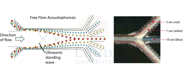
Free Flow Acoustophoresis
Most separation relies on the fact that the primary acoustic radiation forces scales with the volume of the cell, i.e. larger cells move faster towards the pressure node than smaller cells. By tuning the flow rate and flow splitter ratios, it is possible to achieve separation by dividing the outlet flow into different fractions before all cells have reached the pressure node, this mode of operation is called Free Flow Acoustophoresis (FFA). By utilizing the laminar flow present in the microfluidic domain it is possible to separate flow trajectories into multiple outlets.
Petersson et al. (2007) showed microchip FFA in a set-up with four outlets demonstrating discrimination between 2, 5, 8 and 10 μm polystyrene beads.
Multiple biomedical applications have utilized the acoustic multioutlet separation device, including Dykes 2011, Augustsson 2012, Grenvall 2015, and Urbansky 2019.
References
Petersson F. et al., Free Flow Acoustophoresis: Microfluidic-Based Mode of Particle and Cell Separation, Analytical Chemistry, 2007, 79, 5117-5123
Dykes J. et al., Efficient Removal of Platelets from Peripheral Blood Progenitor Cell Products Using a Novel Micro-Chip Based Acoustophoretic Platform, PLoS ONE, 2011, 6, e23074
Augustsson P. et al., Microfluidic, Label-Free Enrichment of Prostate Cancer Cells in Blood Based on Acoustophoresis, Anal Chem, 2012, 84, 7954-7962
Grenvall C. et al., Concurrent Isolation of Lymphocytes and Granulocytes Using Prefocused Free Flow Acoustophoresis, Anal Chem, 2015, 87, 5596-5604
Urbansky A. et al., Label-free separation of leukocyte subpopulations using high throughput multiplex acoustophoresis, Lab Chip, 2019, 19, 1406-1416