Life Science Microfluidics
The Division offers infrastructure and assistance within microfluidics, acoustofluidics, and microfabrication. We can design and develop microsystems for processing of body/bodily fluids, cell sorting, immobilization, and neural electrodes.
Submissions
Submit your project request here.

Examples of available technology and past projects:

Micromilling down to 50 µm. Photolitography down to 5 µm. 3D-printing: Extruder, SLA (400 µm), DLP (10 µm features, 2 µm spots, 5 µm layers). Silicon and glass microfabrication.
Equipment examples: (external links)
Boston Microfabrication MicroArchS230
SLA (stereo lithography) printer
Datron Neo Series 2 (Swedish link)

Gradient generator for Stem Cell regionalization
Modeling neural tube development by differentiation of human embryonic stem cells in a microfluidic WNT gradient, Nature Biotechnology | VOL 38 | November 2020 | 1265–1273.
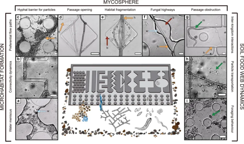
Microfluidic chips provide visual access to in situ soil ecology, COMMUNICATIONS BIOLOGY | (2021) 4:889
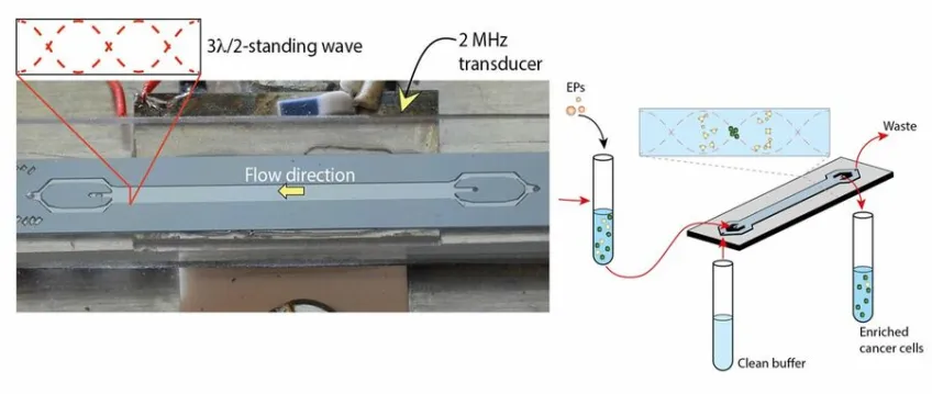
Two-Step Acoustophoresis Separation of Live Tumor Cells from Whole Blood (2021) In Analytical Chemistry 93(51). p.17076.

Acoustic nanoparticle trapping enables isolation and purification of extracellular vesicles in samples as small as e.g. 10 µL blood plasma.

Biogrid—a microfluidic device for large-scale enzyme-free dissociation of stem cell aggregates, Lab Chip, 2011, 11, 3241
Talk to us
Discuss the feasibility of your project with our Project Coordinator:
Martin Bengtsson
martin [dot] bengtsson [at] bme [dot] lth [dot] se (martin[dot]bengtsson[at]bme[dot]lth[dot]se)
Managers


Project Group
Axel Tojo, Martin Bengtsson, Johan Nilsson, Thomas Laurell.